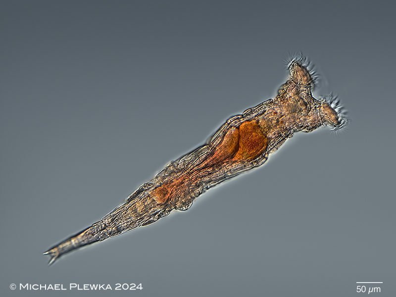
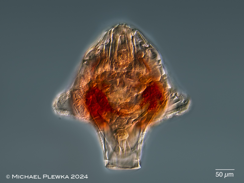
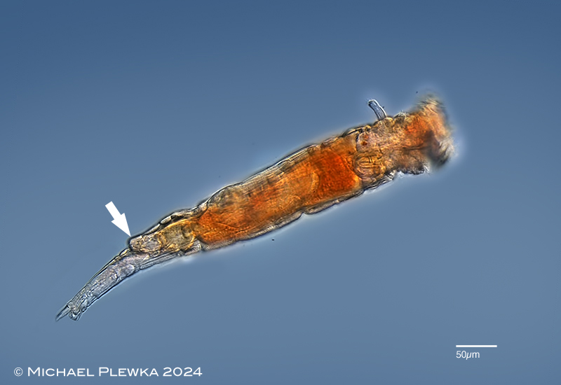
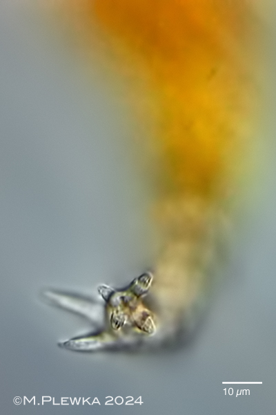
| Philodina cf alata, swimming specimen, dorsoventral view. Because of the orange gut and also reddish-colored integument this species may be easily confused with Philodina roseola. Also visible are the acute spurs. (1). |
| |
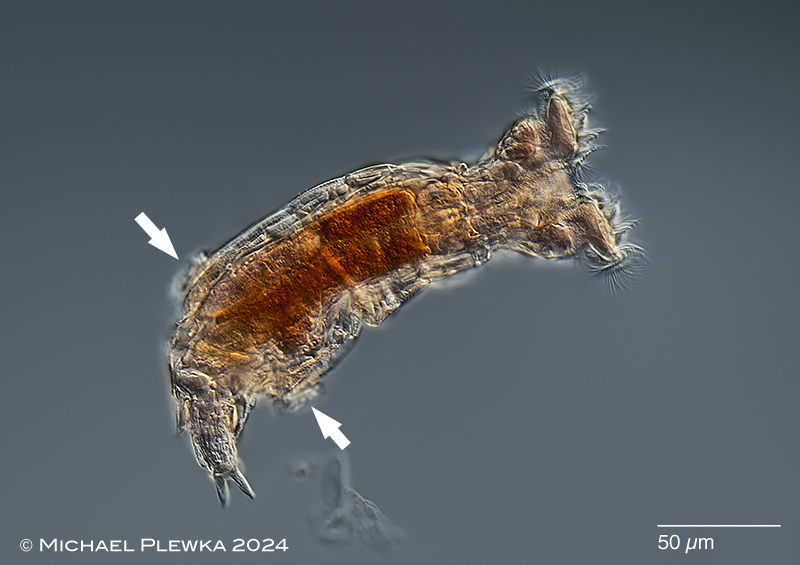 |
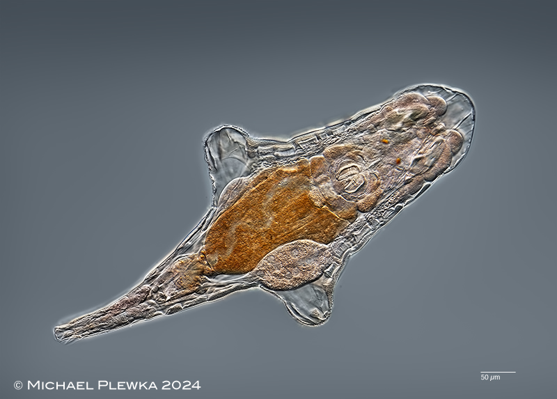
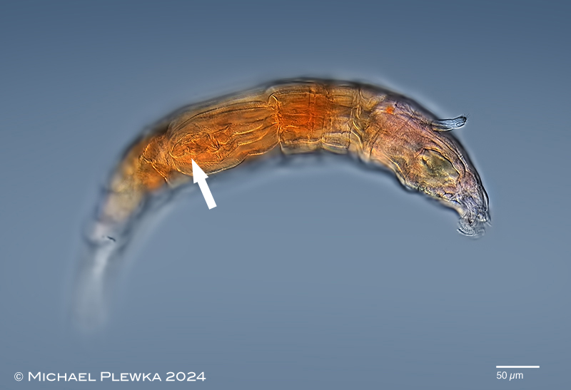
| Philodina cf alata, feeding specimen, ventral view. When feeding the trunk is slightly contracted. In this contraction status the lateral processes become slightly visible (arrows) (1). |
| |
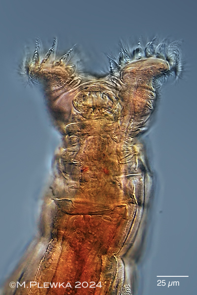 |
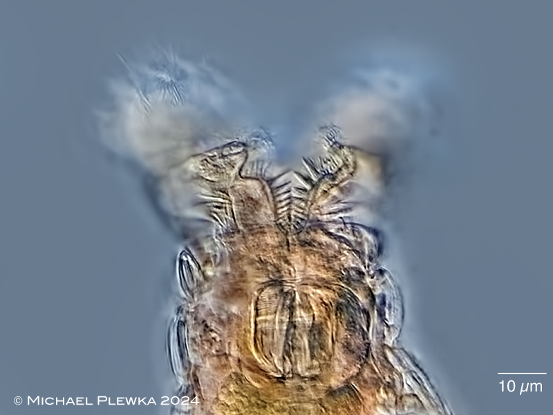
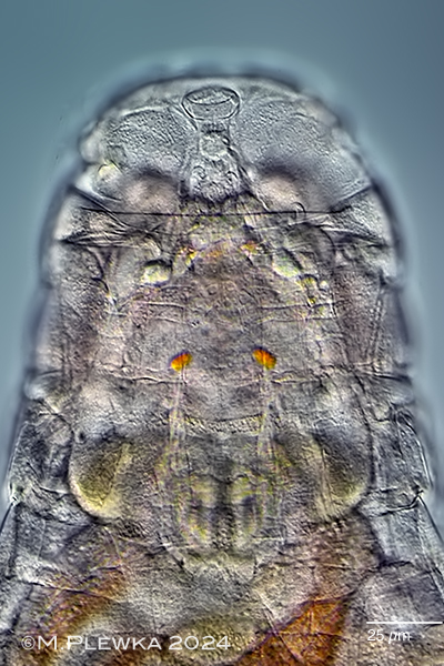
| Philodina cf alata, left: anterior part of swimming specimen with extended corona, dorsal view. Focal plane on the bolsters beween upper lip and trochal columns (pedicels). Optical transect of the rostrum visible). (1). |
| |
| |
 |
 |
 |
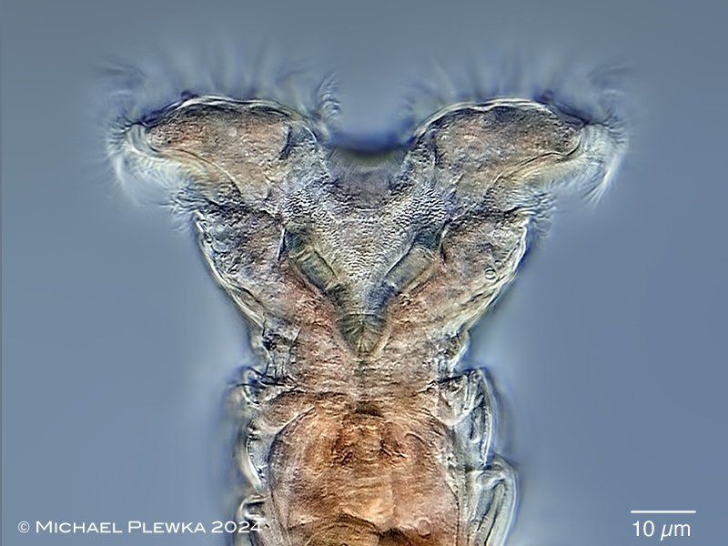 |
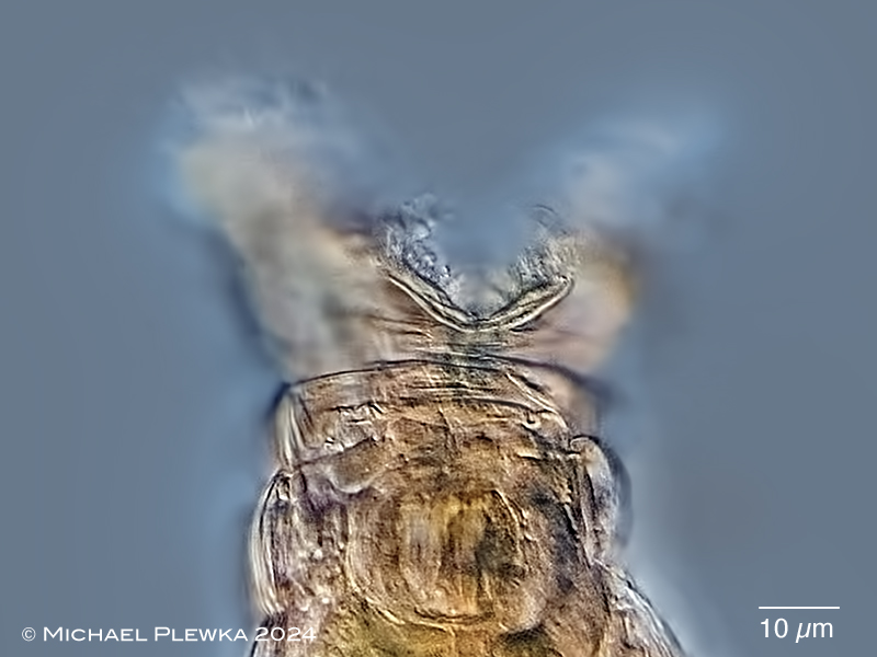
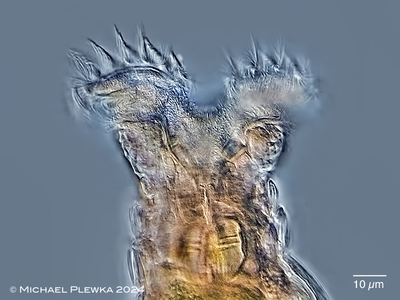
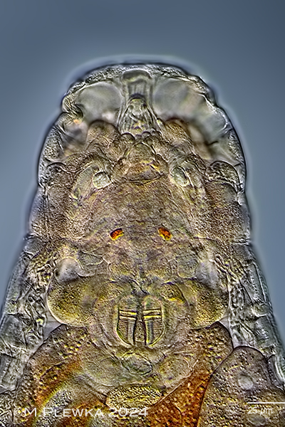
| Philodina cf alata, 4 images of the anterior part in ventral view, different focal planes from ventral to dorsal. 1st: focal plane on the lower lip. 2nd: focal plane on the cilia of the ventral buccal field. 3rd: focal plane on the ciliated field of the trochal columns and the buccal tube (buccal tube length < ramus length). 4th: focal plane on the ciliated field of the trochal columns and the dorsal buccal field. |
| |
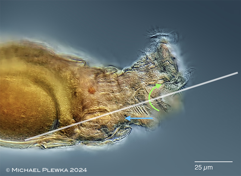 |
| Philodina alata; whirling; lateral view. Focal plane on the cilia of the buccal field/ buccal tube. There are two different pairs of ciliary fields generating different water currents. The arrows indicate the direction of the metachronal waves of these cilia, not the direction of the water flow. Although the direction of the metachronal waves indicated by the green arrow is to the dorsal side, the water flow is to the ventral side. Particles in this area are transported outwards. The blue arrow marks the pair of ciliary fields that create a current towards the mastax. The white line marks the plane of the optical transect no.2 in the upper image sequence. Similar behavior of the coronal cilia could be observed and documented for Philodina roseola. |
| |
| |
| |
| |
 |
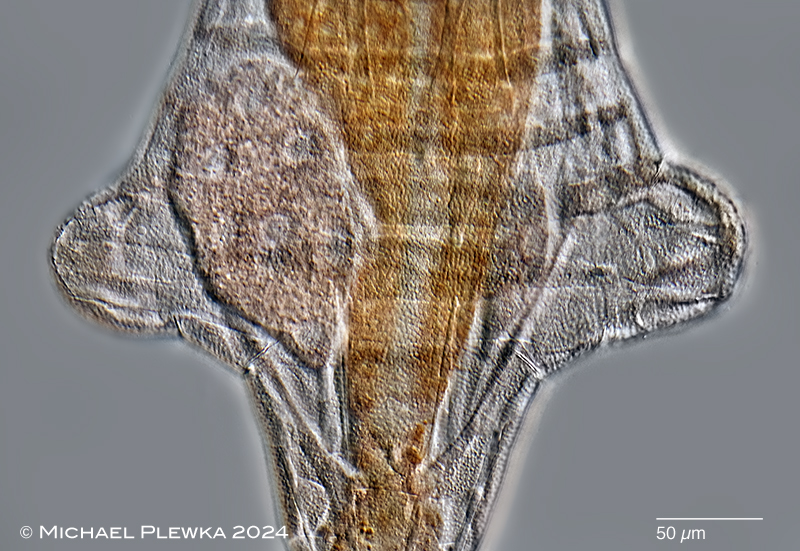
| Philodina cf alata, contracted specimen (corona and foot retracted), dorsal view. (1). |
| |
 |
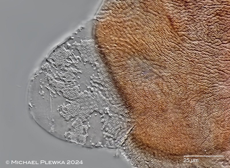
| Philodina cf alata; compressed specimen with retracted corona, showing the two lateral processes. (1). |
| |
  |
| Philodina cf alata; two images of a specimen with retracted corona, slightly compressed by coverslide. Different focal planes. (1). |
| |
 |
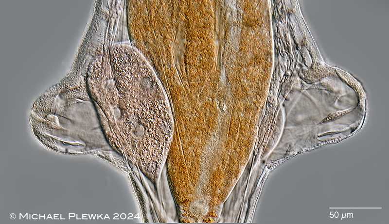 |
| Philodina cf alata; two image sof the same compressed specimen. Upper: focal plane on the integument. Also the transversal muscles are visible due to their optical polarizing properties. Lower: focal plane on the two vitellaria of different size and the muscles in the lateral processess (1). |
| |
 |
 |
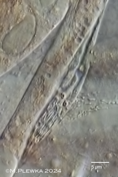 |
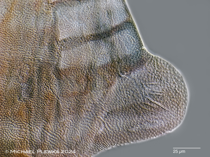
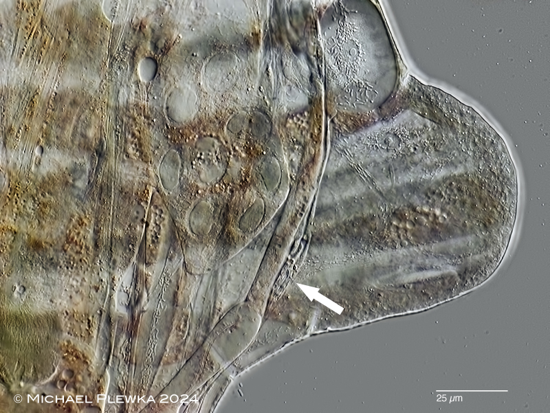
| Philodina cf alata; detail of the right lateral process. Upper image: focal plane on the granulated integument with pores. Lower image: focal plane on the vitellarium (with 8 nuclei). Also visible are some longitudinal and transversal mucles. The arrow points to one of the nephridial organs; a crop can be seen in the lowest image .(1). |
| |
 |
| Philodina cf alata; after maceration with SDS some some structures of the integument of the lateral processes are dissolved. No further structures (i.e. pores ; sensory organs like lateral antennae of monogononts) are visible in the light microscope.(1). |
| |
 |
| Philodina cf alata; lateral view of creeping specimen (i.e. corona is retracted); focal plane on the right lateral process which is contracted and therefore unconspicuous (arrow). Also in focus are the right cervical eyespot, the dorsal antenna and the cilia of the rostrum. (still image from video) |
| |
 |
| Philodina cf alata; lateral view of swimming specimen (i.e. corona is extended); focal plane on the boss on the preanal segment. Also in focus is the dorsal antenna. (still image from video) |
| |
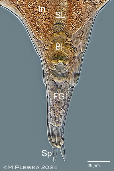  |
| Philodina cf alata, left: posterior part of slightly compressed specimen. Bl: bladder; FG: foot glands; In: intestinum; SL: stomach lumen; Sp: acute spurs. Right: foot with 4 toes and 2 acute spurs (spur length (SL): 15µm) with interspace (2µm); (1). |
| |
| |
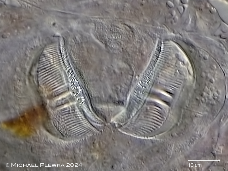 |
| Philodina cf alata;, ramate trophi. cephalic view; with dental formula (DF):2+1/1+2 ; ramus length (RaL): 31µm. |
| |
| Sample courtesy of N. Benvenuty; Spain. |
| |
| |
|
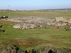
Location (1): Location: Villa del Rey; Cáceres province; Spain, (1) |
 |
| |
| Habitat (1): lithothelma |
| |
| Date: coll.: 01.02.2024; img.: 12.02.2024 |
| |
|
|
|
|
|
| |
| |
| |
|
|
|
|